
Hip
- Anatomy
- Conditions
- Procedures
Hip Anatomy
The hip joint is the largest weight-bearing joint in the human body. It is also referred to as a ball and socket joint and is surrounded by muscles, ligaments, and tendons. The thigh bone or femur and the pelvis join to form the hip joint.
Any injury or disease of the hip will adversely affect the joint's range of motion and ability to bear weight.
The hip joint is made up of the following:
- Bones and joints
- Ligaments of the joint capsule
- Muscles and tendons
- Nerves and blood vessels that supply the bones and muscles of the hip
Bones & Joints
The hip joint is the junction where the hip joins the leg to the trunk of the body. It is comprised of two bones: the thigh bone or femur and the pelvis which is made up of three bones called ilium, ischium, and pubis. The ball of the hip joint is made by the femoral head while the socket is formed by the acetabulum. The Acetabulum is a deep, circular socket formed on the outer edge of the pelvis by the union of three bones: ilium, ischium and pubis. The lower part of the ilium is attached by the pubis while the ischium is considerably behind the pubis. The stability of the hip is provided by the joint capsule or acetabulum and the muscles and ligaments which surround and support the hip joint.
The head of the femur rotates and glides within the acetabulum. A fibrocartilaginous lining called the labrum is attached to the acetabulum and further increases the depth of the socket.
The femur or thigh bone is one of the longest bones in the human body. The upper part of the thigh bone consists of the femoral head, femoral neck, and greater and lesser trochanters. The head of the femur joins the pelvis (acetabulum) to form the hip joint. Next to the femoral neck, there are two protrusions known as greater and lesser trochanters which serve as sites of muscle attachment.
Articular cartilage is the thin, tough, flexible, and slippery surface lubricated by synovial fluid that covers the weight-bearing bones of the body. It enables smooth movements of the bones and reduces friction.
Ligaments
Ligaments are fibrous structures that connect bones to other bones. The hip joint is encircled with ligaments to provide stability to the hip by forming a dense and fibrous structure around the joint capsule. The ligaments adjoining the hip joint include:
- Iliofemoral ligament – This is a Y-shaped ligament that connects the pelvis to the femoral head at the front of the joint. It helps in limiting over-extension of the hip.
- Pubofemoral ligament – This is a triangular shaped ligament that extends between the upper portion of the pubis and the iliofemoral ligament. It attaches the pubis to the femoral head.
- Ischiofemoral ligament – This is a group of strong fibers that arise from the ischium behind the acetabulum and merge with the fibers of the joint capsule.
- Ligamentum teres – This is a small ligament that extends from the tip of the femoral head to the acetabulum. Although it has no role in hip movement, it does have a small artery within that supplies blood to a part of the femoral head.
- Acetabular labrum – The labrum is a fibrous cartilage ring which lines the acetabular socket. It deepens the cavity increasing the stability and strength of the hip joint.
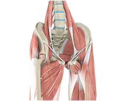
Muscles & Tendons
A long tendon called the iliotibial band runs along the femur from the hip to the knee and serves as an attachment site for several hip muscles including the following:
- Gluteals – These are the muscles that form the buttocks. There are three muscles (gluteus minimus, gluteus maximus, and gluteus medius) that attach to the back of the pelvis and insert into the greater trochanter of the femur.
- Adductors – These muscles are in the thigh which help in adduction, the action of pulling the leg back towards the midline.
- Iliopsoas: This muscle is in front of the hip joint and provides flexion. It is a deep muscle that originates from the lower back and pelvis, and extends up to the inside surface of the upper part of the femur.
- Rectus femoris – This is the largest band of muscles located in front of the thigh. They also are hip flexors.
- Hamstring muscles- These begin at the bottom of the pelvis and run down the back of the thigh. Because they cross the back of the hip joint, they help in extension of the hip by pulling it backwards.
Nerves & Arteries
Nerves of the hip transfer signals from the brain to the muscles to aid in hip movement. They also carry the sensory signals such as touch, pain, and temperature back to the brain.
The main nerves in the hip region include the femoral nerve in the front of the femur and the sciatic nerve at the back. The hip is also supplied by a smaller nerve known as the obturator nerve.
In addition to these nerves, there are blood vessels that supply blood to the lower limbs. The femoral artery, one of the largest arteries in the body, arises deep in the pelvis and can be felt in front of the upper thigh.
Hip Movements
All the anatomical parts of the hip work together to enable various movements. Hip movements include flexion, extension, abduction, adduction, circumduction, and hip rotation.
Conditions
Hip Injuries & Tears
Snapping Hip
The hip is an important joint that helps us walk, run and jump. The ball-and-socket joint in the hip is formed between the round end of the femur (thighbone) and the cup-shaped socket of the acetabulum (part of the hip bone). Joint stability in the hip region is achieved through the labrum (a strong fibrous cartilage), which covers the acetabulum and seals it, and ligaments (tissue connecting bone to bone) and tendons (tissue connecting muscle to bone) that encase the hip and control the hip movements.
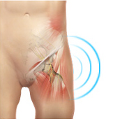
Hip Pain
Hip pain, one of the common symptoms patients complain of, may not always be felt precisely over the hip joint. Pain may be felt in and around the hip joint and the cause for pain is multifactorial. The exact position of your hip pain suggests the probable cause or underlying condition causing pain. Pain felt inside the hip joint or your groin area is more likely to be because of the problems within the hip joint.
Muscle Strains
A tear in the muscle fibers caused by either a fall or direct blow to the muscle, overstretching and overuse injury is called a strain. Muscle strains often occur in the hip region whenever a muscle contracts suddenly from its stretched position. It can be mild, moderate or severe and depends on the level of injury. The chances of having a hip muscle strain becomes high if you have had a previous injury in the area or if there is no warm-up before exercising.

Femoroacetabular Impingement
Femoroacetabular impingement (FAI) is a condition where there is too much friction in the hip joint from bony irregularities causing pain and decreased range of hip motion. The femoral head and acetabulum rub against each other creating damage and pain to the hip joint. The damage can occur to the articular cartilage (the smooth white surface of the ball or socket) or the labral tissue (the lining of the edge of the socket) during normal movement of the hip.
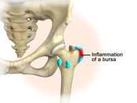
Hip Bursitis
Hip bursitis is a painful condition caused by inflammation of a bursa in the hip. Bursae are fluid filled sacs present in joints between bone and soft tissue to reduce friction and provide cushioning during movement.
The bony prominence of the hip is called greater trochanter and is present on the outer side of the upper thigh bone or femur. The bursa overlying it is called trochanteric bursa. Another bursa is located towards the groin region and is called iliopsoas bursa. Bursitis of the trochanteric bursa is more common than that of iliopsoas bursa.
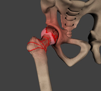
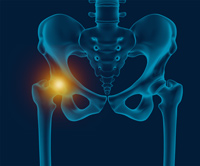
Avascular Necrosis
Avascular necrosis, also called osteonecrosis is a condition in which bone death occurs because of inadequate blood supply to it. Lack of blood flow may occur when there is a fracture in the bone or a joint dislocation that may damage nearby blood vessels. Chronic use of high doses of steroid medications and heavy alcohol consumption are the two main risk factors of avascular necrosis. Initially, small breaks appear in the bone that may eventually collapse.
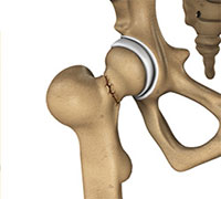
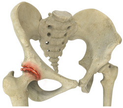
Hip Fracture
The hip joint is a “ball and socket” joint. The “ball” is the head of the femur, or thigh bone, and the “socket” is the cup shaped acetabulum. The joint surface is covered by a smooth articular surface that allows pain free movement in the joint.
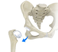
Hip Dislocation
The hip joint is a ball and socket joint. The “ball” is the head of the femur, or thigh bone, and the “socket” is the cup shaped acetabulum. The joint is surrounded by muscles, ligaments, and tendons that support and hold the bones of the joint in place. Hip dislocation occurs when the head of the femur moves out of the socket.
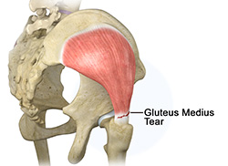
Gluteus Medius Tear
A gluteus medius tear is a condition characterized by severe strain on the gluteus medius muscle that results in partial or complete rupture of the muscle.
The gluteus medius is one of the major muscles of the hip and is essential for movement of the lower body and keeping the pelvis level during ambulation.
Hip Labral Tear
A hip labral tear is an injury to the labrum, the cartilage that surrounds the outside rim of your hip joint socket. The hip joint is a ball and socket joint in which the head of the femur is the ball and the pelvic acetabulum forms the socket. The labrum helps to deepen the socket and provide stability to the joint. It also acts as a cushion and enables smooth movements of the joint.
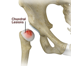
Chondral Lesions or Injuries
The hip joint is one of the largest weight-bearing joints in the body, formed by the thigh bone or femur and the acetabulum of the pelvis. It is a ball and socket joint with the head of the femur as the ball and the pelvic acetabulum forming the socket. The joint surface is covered by a smooth articular cartilage which acts as a cushion and enables smooth movements of the joint. A chondral injury refers to an injury of the articular cartilage, covering the joint.
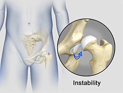
Hip Instability
The hip plays an important role in supporting the upper body weight while standing, walking and running, and hip stability is crucial for these functions. The femur (thigh bone) and acetabulum (hip bone) join to form the hip joint, while the labrum (tissue rim that seals the hip joint) and the ligaments lining the hip capsule maintain the stability of the hip.
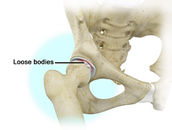
Loose Bodies
Loose bodies are small loose fragments of cartilage or a bone that float around the joint. The loose bodies can cause pain, swelling, locking and catching of the joint. Loose bodies occur if there is bleeding within the joint, death of tissues lining the joints associated with tuberculosis, osteoarthritis, and rheumatoid arthritis. Other causes include fractures, trauma, bone and cartilage inflammation, and benign tumors of the synovial membrane.
Hip Groin Disorders
Hip and groin disorders are more common in athletes, caused by rapid acceleration and deceleration motion.
The rehabilitation time for hip and groin injuries are longer than most other injuries, therefore early and accurate diagnosis is essential. The management of hip and groin injuries is complex due to the presence of multiple anatomic structures in that region.
Hip Distraction
The hip joint is one of the most important and flexible joints in the human body which allows us to walk, run, bend and perform physical activities. It is a ball (femoral head) and socket joint formed between the hip bone and femur (thigh bone). The hip joint is surrounded by strong muscles and tough ligaments that prevent dislocation of the hip.
Subtrochanteric Hip Fracture
A hip fracture is a break that occurs near the hip in the upper part of the femur or thigh bone. The thigh bone has two bony processes on the upper part - the greater and lesser trochanters. The lesser trochanter projects from the base of the femoral neck on the back of the thigh bone. Hip fractures can occur either due to a break in the femoral neck, in the area between the greater and lesser trochanter or below the lesser trochanter.
Hip Abductor Tears
Hip abductors are a major group of muscles found in the buttocks. It includes the gluteus maximus, gluteus medius, gluteus minimus, and tensor fascia lata muscles.
The Gluteus medius arises at the top of the pelvic bone and runs to attach on the outer side of the thighbone or femur. The muscle controls side-to-side movement of the hip and stabilizes hip movement.
Hip Synovitis
Hip synovitis, also called transient hip synovitis or toxic synovitis is a condition in which there is inflammation of the synovial tissues surrounding the hip joint causing hip pain. It is the most common reason for sudden hip pain occurring in young children between the age of 2 and 9. It affects boys more commonly than girls and most of the times, the hip joint on only one side is affected.
Irritable Hip
Irritable hip, also known as acute transient synovitis, is a common disorder of childhood characterized by onset of hip pain and limping. The term transient means that it does not usually last long. It usually occurs before puberty and affects only one hip. Boys aged between 4 to 10 years are most often affected 2 to 4 times more than girls.
Hip Tendonitis
Tendons are strong connective tissue structures that connect muscle to bone. Hip tendonitis is a condition associated with degeneration of the hip tendons. This condition is mainly caused due to strain on the tendons which may be due to overuse, or biomechanical problems.
Hip Pointers
The hip joint consists of 2 bones, the hip bone and the leg bone. An injury or bruise to one of these bones or the surrounding muscles or tissues is termed a hip pointer. This type of injury is mainly caused due to a sudden blow or hit on the hip which can occur from sports activities such as football, rugby, volleyball, basketball, cycling or skating.
Developmental Dysplasia
Developmental dysplasia of the hip (DDH) or Hip dysplasia is a condition which is seen in infants and young children because of developmental problems in the hip joint. The femur (thigh bone) partially or completely slips out of the hip socket causing dislocation at the hip joint. It is most common in first born baby with family history of the disorder.
Legg-Calve-Perthes-Disease
Legg-Calve-Perthes Disease (LCPD) or Perthes disease is a disorder of the hip that affects children, usually between the ages of 4 and 10. It usually involves one hip, although it can occur on both sides in some children. It occurs more commonly in boys than girls.
Slipped Capital Femoral Epiphysis
Slipped capital femoral epiphysis (SCFE) is an unusual disorder of the hip where the ball at the upper end of the thigh bone (femur) slips in a backward direction. This is caused due to weakness of the growth plate. This condition is commonly caused during accelerated growth periods such as the onset of puberty.
Hip Arthritis
Osteoarthritis of the Hip
Osteoarthritis, also called degenerative joint disease is the most common form of arthritis. It occurs most often in older people. This disease affects the tissue covering the ends of bones in a joint (cartilage). In a person with osteoarthritis, the cartilage becomes damaged and worn out causing pain, swelling, stiffness and restricted movement in the affected joint.
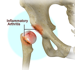
Inflammatory Arthritis of the Hip
Inflammation of the joints is referred to as arthritis. The inflammation arises when the smooth covering (cartilage) at the end surfaces of the bones wears away. In some cases, the inflammation is caused when the lining of the joint becomes inflamed as part of an underlying systemic disease. These conditions are referred to as inflammatory arthritis.
Transient Osteoporosis of the Hip
Transient osteoporosis of the hip is a rare condition that causes bone loss temporarily in the upper part of the thighbone (femur). It is mostly found in young or middle aged men between the ages of 30 and 60, and women in their later stages of pregnancy or early postpartum period (following childbirth). It is characterized by abrupt onset of pain that increases with activity.
Procedures
Non-Surgical Treatments
Hip Injections
Hip joint injections involve injecting medicine directly into the hip joint to diagnose the source of pain or treat pain due to conditions such as arthritis, injury or mechanical stress of the hip joint. Hip pain may be experienced in the hip, buttock, leg or low back. The injection contains a combination of a numbing medicine and cortisone (an anti-inflammatory agent).
Physiotherapy
Physiotherapy or physical therapy is an exercise program that helps you to improve movement, relieve pain, encourage blood flow for faster healing, and restore your physical function and fitness level. The main aim of physical therapy is to make your daily activities, such as walking, getting in and out of bed, or climbing stairs, easier.
Platelet Rich Plasma Therapy
Our blood consists of a liquid component known as plasma. It also consists of three main solid components which include the red blood cells (RBCs), white blood cells (WBCs), and platelets. Platelets play an important role in forming blood clots.
Surgical Treatment
Hip Arthroscopy
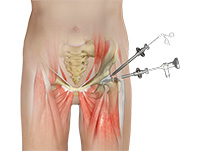
Arthroscopy, also referred to as keyhole or minimally invasive surgery, is a procedure in which an arthroscope is inserted into a joint to check for any damage and repair it simultaneously.
An arthroscope is a small, fiber-optic instrument consisting of a lens, light source, and video camera. The camera projects an image of the inside of the joint onto a large screen monitor allowing the surgeon to look for any damage, assess the type of injury, and repair the problem.
Hip Replacements
- Total Hip Replacement
- Anterior Hip Replacement
- Revision Hip Replacement
- Minimally Invasive Total Hip Replacement
- Outpatient Hip Replacement
- Posterior Hip Replacement
Others
- Hip Implants
- Core Decompression for Avascular Necrosis of the Hip
- Hip FAO Surgery
- Hip Hemiarthroplasty